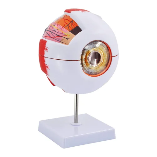
This enlarged eye anatomy model is a highly detailed educational tool designed to help students, trainees, and educators explore the complex structure of the human eye with clarity and precision. Enlarged six times its actual size, the model provides a clear and accurate representation of the major internal and external components of the eye, making it ideal for science classrooms, biology laboratories, medical training institutes, and health education programs. Its large scale allows learners to easily study structures that are normally difficult to observe in real specimens, enabling a deeper understanding of visual anatomy and physiological functions. The model is crafted with attention to anatomical accuracy, showcasing parts such as the cornea, iris, lens, retina, sclera, choroid, optic nerve, and surrounding tissues, all color-coded for easy identification and proper conceptualization of the layered organization of the human eyeball.
Description
The Human Eye Model is a detailed anatomical teaching aid designed specifically for educational and training purposes. It provides a clear three-dimensional representation of the human eye, allowing students to study its structure, function, and role in vision with greater clarity. As a result, learners can move beyond textbook diagrams and gain a deeper understanding through hands-on exploration.
Schools, colleges, medical institutes, and biology laboratories across India widely use human eye models for anatomy education. In addition, educational institutions in countries such as the United Kingdom, South Africa, and Kenya rely on eye models to support interactive learning in biology, medical, and paramedical courses.
Human Eye Model for Interactive Anatomy Learning
The Human Eye Model separates into multiple detachable parts, which allows students to examine each structure individually. This design supports interactive learning by helping users visualize how different components work together to enable vision. Therefore, learners can clearly understand the relationship between the cornea, lens, retina, optic nerve, and other internal structures.
By physically assembling and disassembling the model, students gain insight into light refraction, image formation, depth perception, and the transmission of nerve signals from the retina to the brain. Moreover, this practical engagement improves spatial understanding and long-term concept retention.
Human Eye Model for Vision and Clinical Concept Demonstration
The Human Eye Model plays an important role in explaining both normal vision and common eye disorders. Educators use the model to demonstrate conditions such as myopia, hyperopia, cataracts, glaucoma, and retinal disorders. Consequently, students can connect anatomical structure with functional and clinical outcomes.
The three-dimensional format helps learners visualize internal eye structures more effectively than flat charts or illustrations. As a result, the model supports clearer explanations in both basic biology lessons and advanced medical training sessions.
Stable Stand and Durable Educational Design
The model includes a stable, removable stand that provides firm support during demonstrations. This feature allows instructors to display the model clearly on desks or lab tables, ensuring good visibility for all learners. Additionally, the durable construction withstands frequent handling during classroom and laboratory use.
Suitable for Education and Training Use
This human eye model is suitable for school biology classes, medical and nursing colleges, paramedical institutes, ophthalmology training, anatomy laboratories, and science exhibitions. It aligns well with curricula focused on human anatomy and physiology.
Applications
- Teaching human eye anatomy
- Demonstrating vision and light refraction
- Studying common eye disorders
- Medical and paramedical education
- Classroom and laboratory demonstrations
other educational working models and laboratory kits to enhance hands-on learning in engineering and technical laboratories. In addition, institutions may also explore complementary learning solutions such as Jaadui Pitara educational learning kits to support interactive and concept-based education.