The permanent slide of the transverse section (T.S.) of a monocot stem is an indispensable educational and laboratory resource designed to offer a detailed microscopic view of the anatomical structure of monocotyledonous plant stems. Crafted with precision, this high-quality slide is an essential teaching aid for botany classes, biological science labs, and agricultural studies, enabling students and researchers to closely examine the unique features that differentiate monocot stems from their dicot counterparts. Mounted on a standard glass slide and sealed for long-term preservation, this specimen allows repeated use without degradation, making it a reliable and cost-effective addition to any biological slide collection.
Description
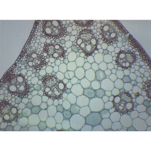
Under the microscope, the T.S. of monocot stem reveals a distinctive anatomical layout, including scattered vascular bundles embedded in a ground tissue matrix. Unlike dicot stems where the vascular bundles are arranged in a ring, monocot stems display an atactostele pattern—an important identifying feature that students can easily observe and analyze using this slide. Each vascular bundle is typically oval or circular in shape and consists of xylem and phloem tissues, often surrounded by a bundle sheath of supportive sclerenchyma cells. The absence of a clearly defined cortex and pith is another key characteristic visible on this slide, helping learners differentiate between the organizational structures of various plant types.
The slide offers an excellent opportunity to observe the parallel arrangement of vascular tissues and the lack of vascular cambium, which explains why monocot stems do not typically undergo secondary growth. This visual understanding aids in grasping fundamental concepts in plant biology, such as the mechanisms of transport and support in monocots. The permanent staining used in the preparation of this slide ensures that all cellular structures—like the xylem vessels, sieve tubes of the phloem, and surrounding parenchymatous ground tissue—are clearly distinguishable under light microscopy. The slide also allows observation of the outer epidermal layer, which may feature a cuticle for protection and may include scattered stomata in certain species.
Designed to meet academic and research needs, the permanent slide of T.S. of monocot stem is highly beneficial for secondary school biology labs, undergraduate botany courses, and advanced plant anatomy studies. Its clear labeling and professionally prepared sample make it ideal for classroom demonstrations, microscopy practice, practical exams, and independent student exploration. Teachers and instructors will find it especially useful in explaining plant structure, tissue differentiation, and comparative anatomy between monocots and dicots, offering visual reinforcement to theoretical content.
Furthermore, this slide is a valuable tool in fields such as horticulture, agronomy, and forestry, where understanding plant internal structure is essential for applications related to crop improvement, disease identification, and plant breeding. The permanence of the specimen ensures it retains clarity over time, resisting damage from regular handling or environmental factors. Its consistent quality and durability make it a preferred choice for institutions aiming to build a long-lasting and reliable collection of botanical teaching materials.
In conclusion, the permanent slide of the transverse section of a monocot stem serves as a vital educational and diagnostic tool. With its scientifically accurate presentation, excellent clarity, and durable build, it supports a wide range of academic and practical applications. Whether used for instruction, research, or examination purposes, this slide is an invaluable resource for anyone studying or teaching plant biology and anatomy.