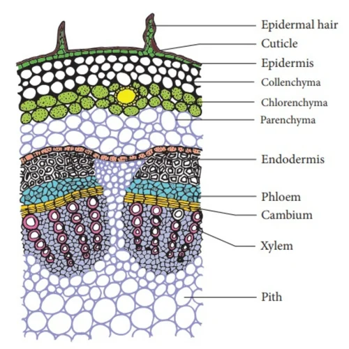
The permanent slide of the transverse section (T.S.) of a dicot stem is an essential educational tool designed to provide a clear, detailed view of the internal anatomical structure of dicotyledonous plant stems. Ideal for biology classrooms, botanical laboratories, and academic institutions, this professionally prepared microscope slide is a staple in the study of plant anatomy, supporting lessons in vascular tissue differentiation, primary and secondary growth, and plant physiology. The T.S. of dicot stem slide typically features clearly identifiable tissues such as the epidermis, cortex, endodermis, pericycle, vascular bundles with xylem and phloem, and centrally located pith. These features are distinctly stained and permanently preserved, allowing repeated and reliable observations under a compound microscope.
Description
One of the key highlights of this permanent slide is the arrangement of vascular bundles in a ring, which is characteristic of dicot stems. This ring-like structure is an important anatomical marker distinguishing dicots from monocots. The xylem and phloem tissues within these bundles are easily identifiable, with the xylem facing the inner side and the phloem positioned toward the outer side. Between these tissues lies the cambium, which plays a vital role in secondary growth and wood formation. This arrangement is fundamental to understanding how dicot plants grow and transport water, minerals, and food throughout their bodies. The permanent nature of this slide ensures that these intricate structures remain intact and visible over long periods, providing a consistent and accurate reference for students and researchers alike.
The preparation process of this slide includes precise sectioning and staining using reliable techniques to highlight cell walls, vascular tissues, and other cellular components. The result is a high-contrast, well-preserved image that brings the internal organization of a dicot stem to life under the microscope. This slide offers an immersive learning experience by allowing close examination of plant tissue layers, including the protective epidermis, the multi-layered cortex involved in storage and support, and the endodermis that regulates the movement of substances into the vascular cylinder. The central pith is also visible, aiding in the identification of the stem’s structural balance and storage functions.
For educators, this permanent slide of T.S. of dicot stem serves as an invaluable resource that enhances classroom demonstrations, practical exams, and laboratory activities. It bridges the gap between theoretical botanical concepts and hands-on microscopy, making abstract ideas more accessible and comprehensible for students. The clarity and durability of the slide ensure that it can be used across multiple academic sessions without the need for repeated sample preparation, saving both time and resources while maintaining educational quality. Additionally, the slide is mounted on a standard-size glass plate with clear labeling and sealed to prevent deterioration, ensuring long-term usability and ease of handling.
In the context of plant taxonomy, physiology, and environmental science education, this slide plays a critical role in fostering a deeper understanding of plant structure and function. It supports curriculum requirements at secondary, undergraduate, and postgraduate levels, and is widely used in botany, agriculture, and forestry studies. The permanent slide of the transverse section of dicot stem is not only a practical teaching aid but also a valuable asset for anyone interested in the detailed anatomy of plants.