The permanent slide of Aspergillus is an essential tool in the study of mycology, microbiology, and general biological sciences. Meticulously prepared using advanced microscopic slide preservation techniques, this slide captures the structural intricacies of Aspergillus, a genus of filamentous fungi that is widely studied for both its industrial importance and medical relevance. The specimen on the slide is stained to highlight the unique morphological features of Aspergillus, such as its septate hyphae, conidiophores, vesicle, and chains of conidia, offering learners and researchers a clear and detailed visualization of its asexual reproductive structures. This high-quality slide enables consistent and accurate observation, making it ideal for use in educational institutions, research laboratories, and training programs.
Description
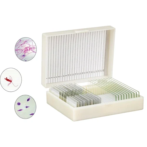
Encased in a high-transparency glass mount, the permanent slide of Aspergillus is designed to provide lasting durability and protection against environmental degradation or accidental damage. The specimen is sealed securely within the slide to prevent contamination and preserve its structural integrity over time. This ensures that the slide remains usable for repeated observations under standard optical or compound microscopes. The clear labeling and precise staining techniques employed in the preparation make it easier for students and educators to identify and study the morphological traits of Aspergillus species, fostering a better understanding of fungal taxonomy and anatomy.
Aspergillus is commonly found in soil, decaying vegetation, and indoor environments, and includes both non-pathogenic and pathogenic species. Its relevance spans various domains such as food spoilage, industrial fermentation, and medical pathology. Studying Aspergillus through a permanent microscope slide allows learners to appreciate its complex reproductive mechanisms, spore formation, and structural adaptations. It is particularly useful in demonstrating the characteristic radial arrangement of conidia and the brush-like structure that is typical of Aspergillus under the microscope. Such clarity in visual representation is invaluable in biology curricula focused on fungi, microbial life, and environmental biology.
The slide also serves as an important diagnostic aid in medical mycology, where accurate identification of fungal morphology plays a critical role in understanding fungal infections like aspergillosis. For healthcare professionals, microbiology students, and laboratory technicians, this permanent slide provides a reference point for comparing clinical isolates with textbook morphology, thus enhancing diagnostic skills and theoretical understanding. Its utility extends to veterinary microbiology and environmental science studies as well, making it a versatile component of any academic or clinical slide set.
In addition to its educational and diagnostic applications, the permanent slide of Aspergillus is a cost-effective and eco-friendly option for institutions aiming to reduce the need for repeated specimen preparation. Unlike temporary mounts, this slide does not require rehydration or special storage conditions, significantly reducing maintenance costs and preparation time. Its robust construction supports safe handling by students, making it suitable even for high school laboratories and undergraduate courses where repeated use and durability are key considerations.
Overall, the permanent slide of Aspergillus is a scientifically valuable, long-lasting, and educationally enriching product that supports a wide range of learning outcomes and research needs. Its detailed visualization of fungal morphology, compatibility with standard microscopy tools, and resilience to frequent use make it an indispensable asset for studying one of the most significant genera of fungi.